Shoulder pathology can be debilitating if the patient does not seek help that can really alleviate the problem. These days, most surgery is done arthroscopically..
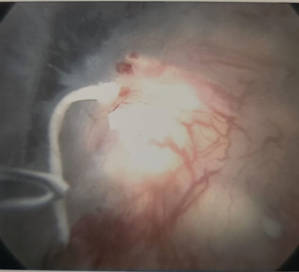
One common problem is a tear of the capsule where it adheres to the shoulderblade part of the joint. This is called a Bankart lesion and can be to the front or the back of the joint. Repair is done arthroscopically using anchors. The outcome of this method is usually very good. Recovery takes about six weeks with the physio being continuously involved.
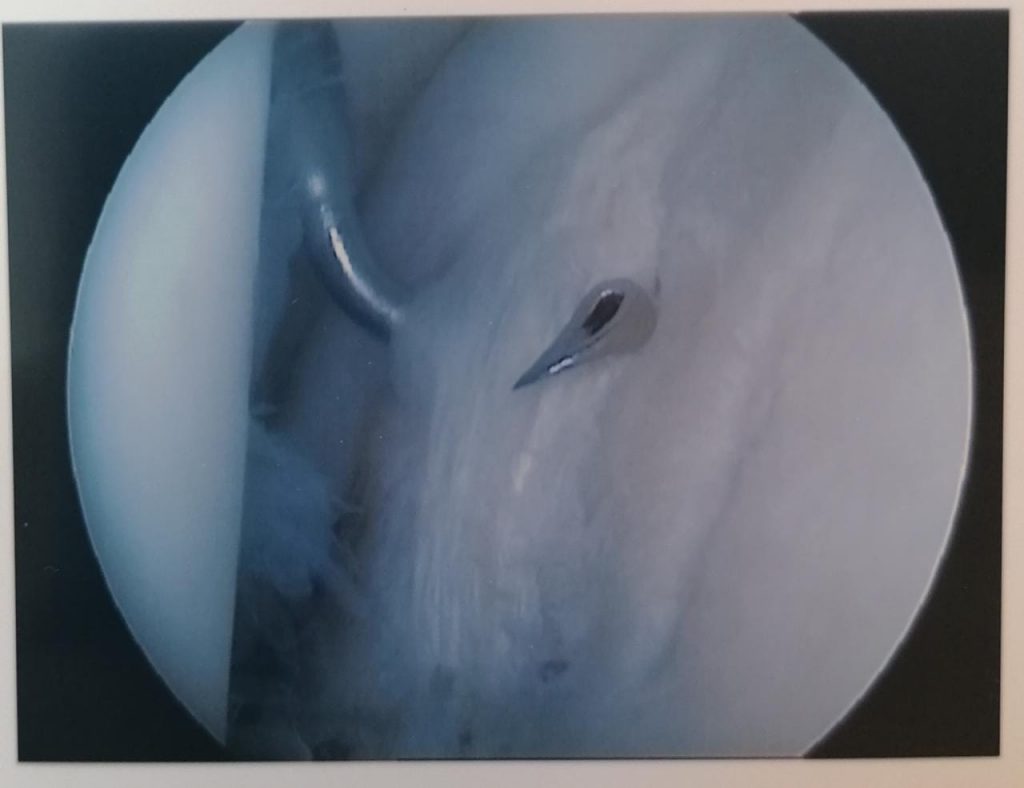
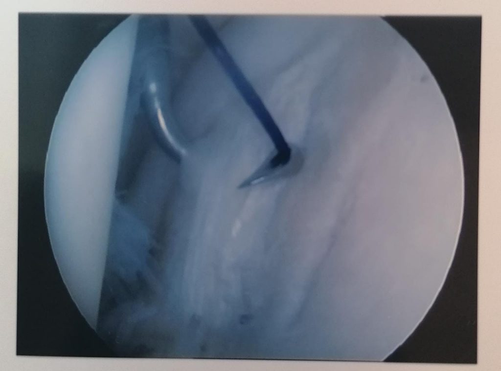
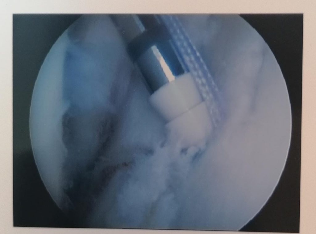
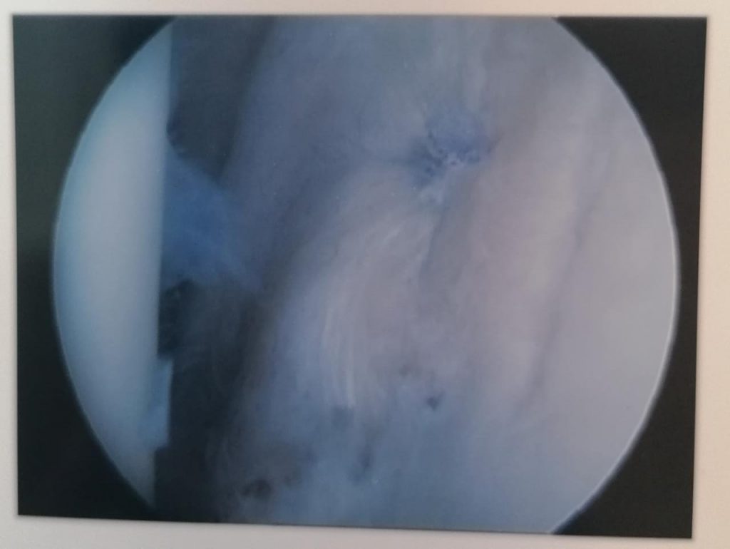
Above are pictures showing the placement of a suture through the edge of the capsule (labrum) to the placement of the anchor as well as the end result. All of this happens through two small incisions.
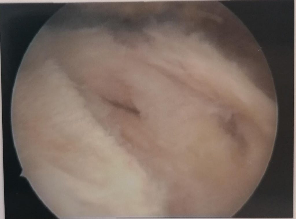
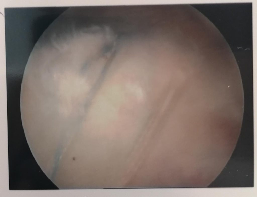
ROTATOR CUFF TEAR
The rotator cuff is the confluence of four muscles that envelop the head of the humerus. It is responsible to initiate motion and to keep the humeral head centered in the glenoid. Age or trauma can cause tears of the rotator cuff, leading to pain and loss of function. Luckily it can be repaired arthroscopically with excellent results
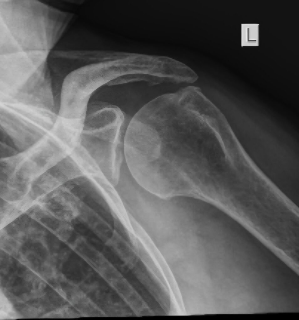
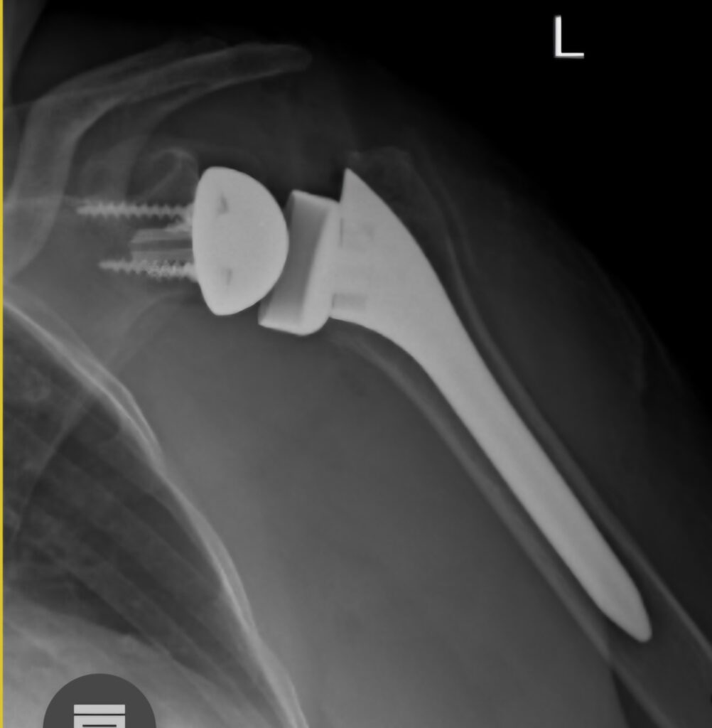
REVERSE TOTAL SHOULDER REPLACEMENT
The rotator cuff is responsible for centering the humeral head in the glenoid and to initiate movement of the shoulder. After initiation the movement is powered by large muscles like the Deltoid and the Pec major etc. If the cuff is torn the first prize is a proper repair , but in patients older than 75 the outcomes are poor. In this patient group there is an alternative, namely a reverse total shoulder replacement. By reversing the ball and socket from the anatomical arrangement, the Deltoid muscle is able to initiate and complete motion of the shoulder. This is and excellent answer to a sticky problem. Not a good option for young patients though
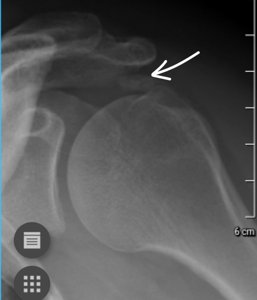
CALCIFIC TENDONITIS
In some patients the rotator cuff calcifies. This is a painless process. Once the calcium deposit starts to dissolve it becomes very painful. Treatment consists of painkillers, hot packs, steroid injection and, if conservative measures fail, surgery. At the time of surgery the calcific deposit is poked with a needle and the liquefied calcium drains. Usually very effective to manage the pain, though not always instantaneous.